|
TO ALL PARTICIPANTS
We had a great workshop this year which was held at the Harvard Medical School in conjunction with the MICCAI 2014 conference. A great day with great presentations and two excellent invited talks by Prof David Haynor and Prof Nassir Navab. Thanks everyone for making this workshop a success and see you next year at CSI 2015! Jack, Ben, Tobi, Shuo PS: The slides of Prof David Haynor's presentation are available here. 
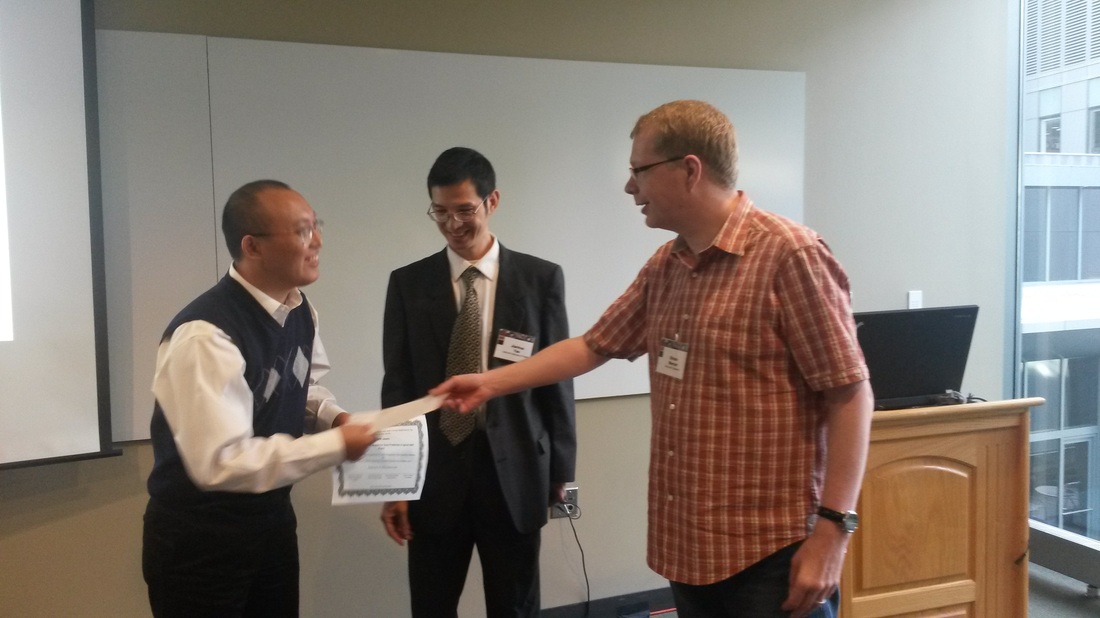
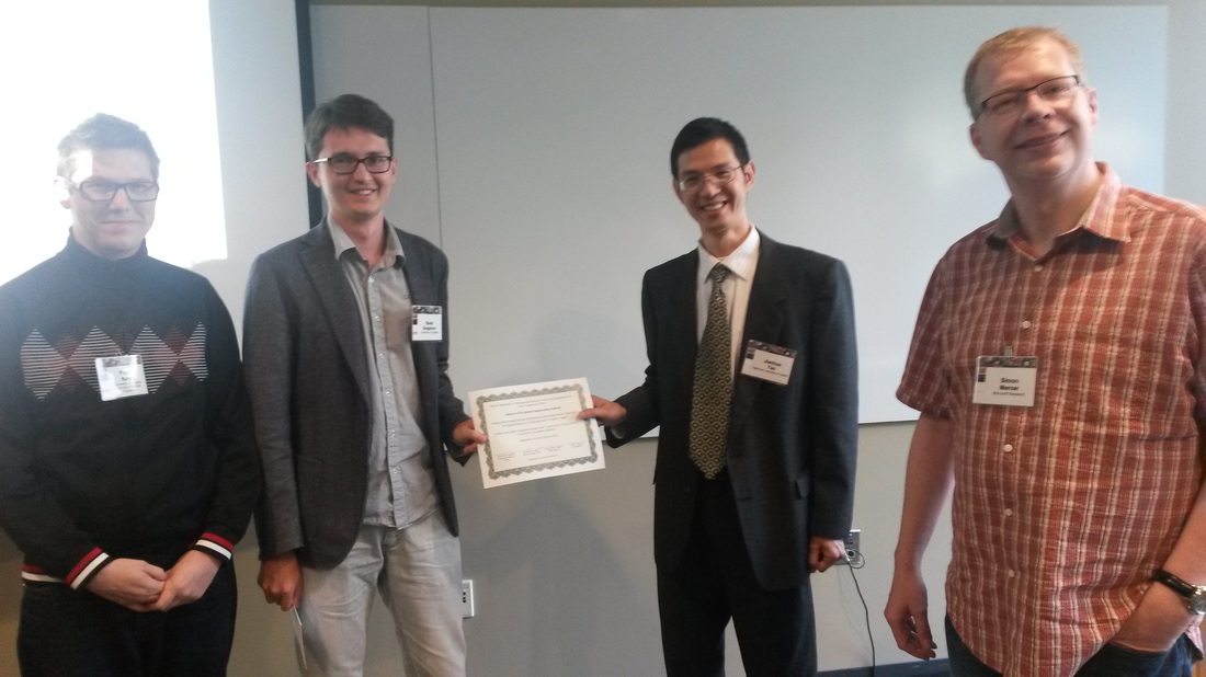
Winners of the Workshop Awards
This year's awards were sponsored by Microsoft Research. We had two categories, 'Winner of Vertebra Segmentation Challenge' and 'Best Workshop Paper'. And the winners are... Best Workshop Paper 2014
Winner of Vertebra Segmentation Challenge 2014
CSI 2014 WORKSHOP
The spine represents both a vital central axis for the musculoskeletal system and a flexible protective shell surrounding the most important neural pathway in the body, the spinal cord. Spine related diseases or conditions are common and cause a huge burden of morbidity and cost to society. Spine imaging is an essential tool for assessing spinal pathologies and there is need for advanced computerized methods that support the physician in diagnosis, therapy and intervention. Venue The 2nd Workshop on Computational Methods and Clinical Applications for Spine Imaging (CSI 2014) is held in conjunction with MICCAI 2014 in Boston. The workshop will be held on Sunday, September 14 at the Joseph B. Martin Conference Center at Harvard Medical School (HMS), 77 Avenue Louis Pasteur. The workshop will be in conference room 214.There will be buses between Kresge Auditorium at MIT and the Martin Conference Center at HMS in the morning (departing at 7:00am and at 7:30am) and in the evening (departing at 5:15pm). Alternatively, you can use public transportation or taxi. CSI 2014 is an event supported by the SpineWeb Initiative. There will be invited talks from two internationally recognized experts in spinal imaging. Prof. David R Haynor and Prof. Nassir Navab will share their expertise and knowledge in the field of clinical spinal applications. Scope of the Workshop We are inviting researchers to submit papers on computational methods in the area of spine imaging and image analysis. Topics of interest include, but are not limited to:
Challenges An important aspect of this year’s workshop is to include computational challenges. Researchers are encouraged to evaluate novel and existing methods on two important image analysis tasks. The first challenge concerns full vertebrae segmentation, the second challenge is on vertebrae localization and identification. For both challenges a set of annotated training data is made available (~20 for segmentation, ~200 for localization/identification) through the SpineWeb Initiative. This data is intended to be used for development, parameter learning, and self-evaluation. Evaluation datasets will be released for both challenges. Participants are asked to run their methods on this data and submit their results which will be evaluated and ranked by the organizers. Challenge results will be presented and discussed during the workshop. |

|
 |
|